3D Ultrasound: Ultrasound technology has changed medical diagnostics, particularly in obstetrics, which is pivotal in monitoring the development of a fetus. The 3D Scan is one of the most advanced ultrasounds among others in offering detailed & accurate imaging.
The technology of 3D Sonography provides a three-dimensional view of the body’s internal structures that helps both healthcare professionals and patients with valuable insights compared to 2D Scans.
In this ultimate article, we will explore the details of 3D ultrasound scans, which cover their purpose, procedure, benefits, constraints, differences between 2D, 3D, and 4D Ultrasounds, and potential applications in healthcare.
- What is a 3D Ultrasound Scan in Pregnancy?
- How Does a 3D Ultrasound Work?
- Different Applications of 3D Scans in Pregnancy
- Advantages of 3D Pregnancy Ultrasound Scans
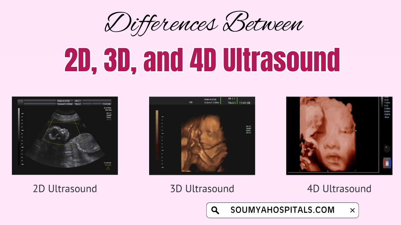
- Differences Between 2D, 3D, and 4D Ultrasound Scanning
- Limitations of 3D Scanning During Pregnancy
- FAQs On 3D Imaging Technology Ultrasound Scan
What is a 3D Ultrasound Scan in Pregnancy?
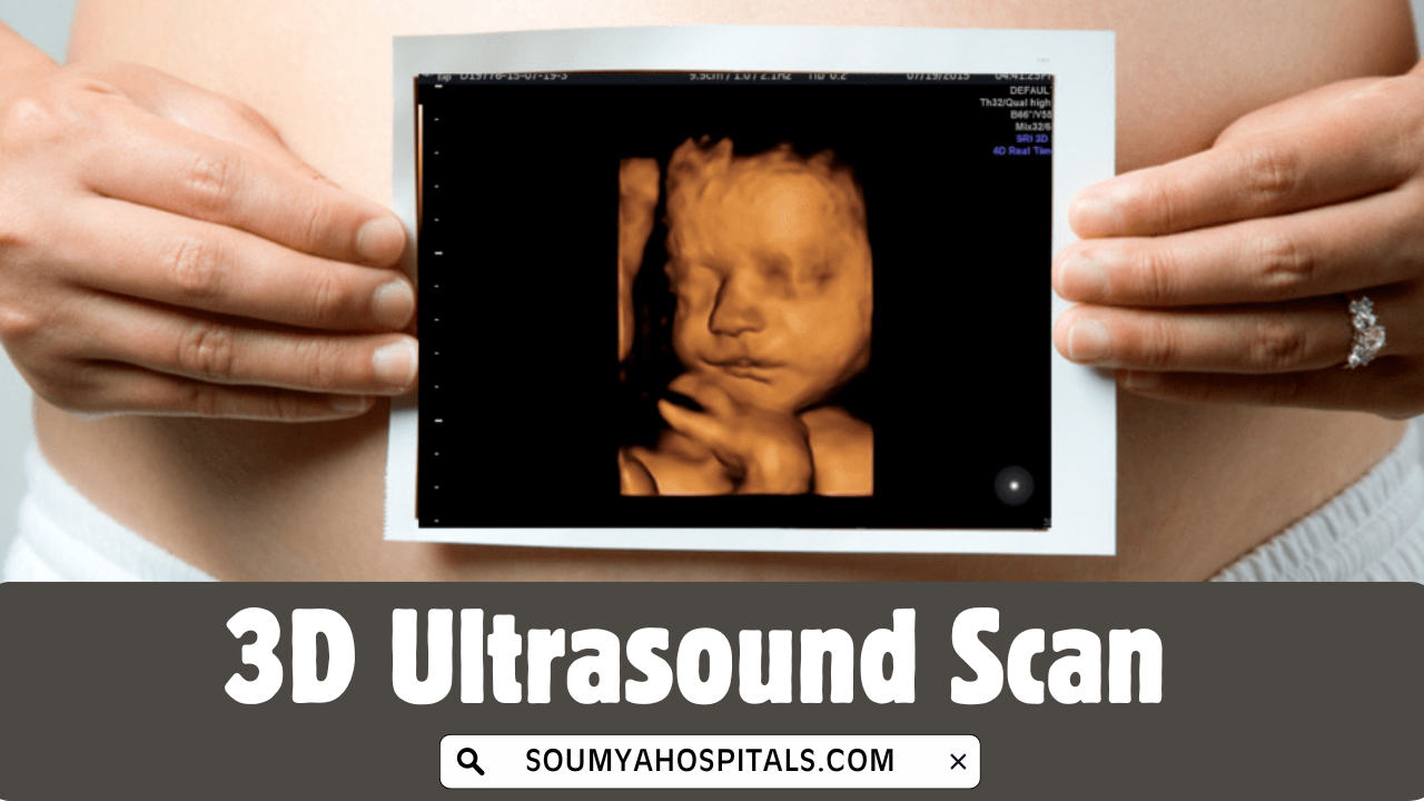
A 3D ultrasound scan produces three-dimensional pictures of the internal organs or fetus in a clear view unlike what is captured by a 2D Scan. Generally, 2D ultrasound scans allow flat, two-dimensional images in black and white, whereas 3D ultrasound authorizes giving a more realistic view, and visualizes shapes & contours easily.
The technology seizes a series of two-dimensional images and renders them into 3D pictures. Like 2D, 3D ultrasound uses the same high-frequency sound waves, but the major difference is seen in the presentation and processing of images.
Most importantly, the three-dimensional pictures of the baby’s organs and fetus assist in diagnosing and monitoring requirements that may need more comprehensive visualization.
What is the Procedure for a Three-Dimensional 3D Pregnancy Scan? | How Does a 3D Ultrasound Work?
Want to be aware of the 3D ultrasound process performed on the mom-to-be? You’re on the right page. Here we discuss the step-by-step 3D ultrasound scan procedure which is quite similar to the 2D ultrasound in pregnancy and differs in image-processing techniques and equipment.

Here is a step-by-step overview of the 3D Pregnancy Scan Process:
Preparation: During the abdominal ultrasound, the patient has to drink water to fill the bladder. It gives clear images of the internal organs and fetus structure by pushing the intestines out of the way.
Application of Gel: After entering the sonography room, the examiner applies a water-based gel to the area where the ultrasound probe, or transducer, will be utilized. It sends the sound waves into the body and ensures the ultrasound works efficiently.
Use of the Transducer: When you get an ultrasound, the sonographer will use a transducer tool to send sound waves into your body. The transmitted sound waves bounce off your insides, including a baby if you're pregnant, and generate a picture of what's inside.
Image Formation: As the sound waves are reflected, they are captured by the ultrasound machine, which processes them to create detailed 3D images. The process takes a few minutes longer than a standard 2D ultrasound since more images are captured and compiled to form the three-dimensional representation.
Analysis: Once the images are compiled, the doctor can analyze them to assess the patient’s condition. For expectant mothers, the images provide a clearer view of the baby’s facial features, limbs, and organs.
Different Applications of 3D Scans in Pregnancy
3D ultrasound scan is a flexible imaging scan in pregnancy and it is utilized in diverse medical fields. Also, it has a wide range of applications in diagnosing and monitoring medical requirements such as:
- Obstetrics and Gynecology
- Musculoskeletal Imaging
- Cardiology
- Oncology
Do Refer: Pregnancy Ultrasound Scan List
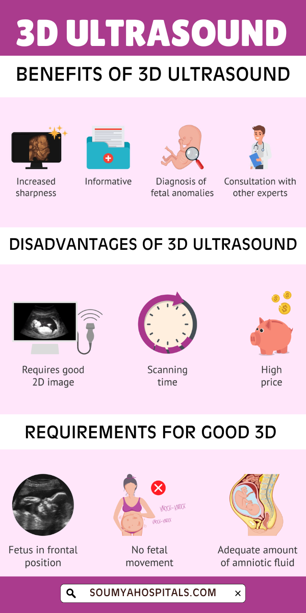
Advantages of 3D Pregnancy Ultrasound Scans
There are numerous benefits of three-dimensional ultrasound scans to both healthcare providers and patients. Some of the key benefits are:
- Better Visualization: First and foremost, the 3D scan is beneficial in offering a detailed visualization of the internal anatomy. It helps healthcare providers to examine the internal organs and fetus condition in clear & accurate pictures which helps to prevent and treat any complication during pregnancy with effective diagnosis.
- Early Detection of Abnormalities: 3D ultrasound scans of pregnancy exhibit more information which helps in identifying the physical problems early. Along with that, midwives can easily find the problems with the child at an early stage and plan to get ready for treatments or surgeries if necessary.
- Slightly Invasive and Safe: Like traditional ultrasound, 3D ultrasound is non-invasive and does not use harmful radiation, making it a safe option for both diagnosis and monitoring. This is particularly important in pregnancy, where the safety of the fetus is a top priority.
- Help in Surgical Planning: In some cases, 3D ultrasound scans can provide valuable information for surgical planning. By offering a clear view of the affected area, doctors can plan more precise and effective surgical interventions.
- Improved Bonding Experience for Expectant Parents: For expectant parents, a 3D ultrasound provides a more tangible and realistic image of their unborn baby. The ability to see the baby’s face and movements in such detail often strengthens the emotional bond between parents and the child.
Differences Between 2D, 3D, and 4D Ultrasound Scanning

Understanding the differences between the various types of ultrasound scans is important when considering their applications.
2D Ultrasound: This is the standard form of ultrasound, producing flat, black-and-white images. It is widely used in routine medical check-ups and pregnancy monitoring. While it is effective for general observation, it lacks the depth and detail provided by 3D ultrasound.
3D Ultrasound: As discussed, 3D ultrasound creates three-dimensional images that offer more detailed views of the internal organs or fetus. It is useful when more in-depth analysis is required.
4D Ultrasound: The term “4D” refers to the addition of real-time movement to 3D images, effectively creating a live video feed. This technology is mostly used in obstetrics, allowing parents to see their baby’s movements in real time.
Limitations of 3D Scanning During Pregnancy
When there are advantages, you can even see minor disadvantages of any test or scan in pregnancy. So remember to take a look at the 3D ultrasounds downsides from below:
- 3D ultrasound cost is more expensive than 2D ultrasound, so patients should prepare mentally about pricing.
- They are not always needed for regular check-ups, as 2D ultrasounds can often do the job.
- Based on various factors like the baby's position or the amount of fluid around the baby, the image quality gets affected, so they might not always be very clear.

FAQs On 3D Imaging Technology Ultrasound Scan
1. What week is best for a 3D baby scan?
The ideal time or best weeks for 3D scans in pregnancy is typically between week 26 and week 32 of pregnancy. In this stage of pregnancy, the 3D ultrasound shares the pictures of the developed baby's facial features.
2. How much does a 3D pregnancy scan cost in the UK Country?
The approximate cost of a 3D Baby Scan in the UK is £90.
3. Is a 3D ultrasound scan safe for a baby?
Yes, 3D scanning during pregnancy is the safest pregnancy test like 2D Scans and other pregnancy ultrasound scans as it performs using sound waves under trained medical practitioners.
Conclusion
3D ultrasound may become more widespread as technology advances, offering even greater insights into the body’s internal workings and improving patient care across various medical fields. Also, a 3D ultrasound scan is a powerful and useful tool that offers detailed and precise imaging of a baby’s internal organs.
For more about pregnancy week by week ultrasound scan list or tests during pregnancy, visit our website @soumyahospitals.com regularly.
